Cone Dystrophy without Rod involvement
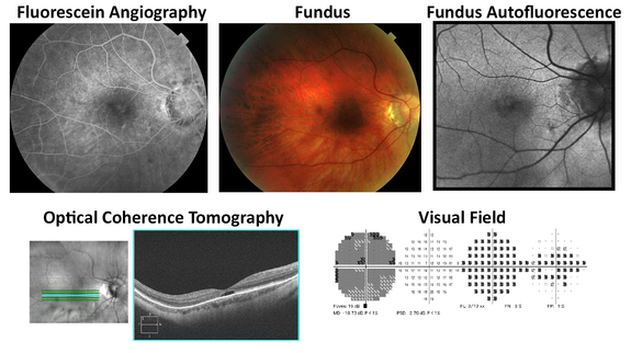
The photos above illustrate cone dystrophy without rod involvement. Only the right eye is shown, but the left eye was similar. The patient was a 69 year old man. His symptoms included reduced visual acuity, incapability of clear vision in bright light (hemeralopia), and the inability to see or distinguish colors (color blindness). The condition seen here was x-linked recessive; his maternal uncle had a similar condition. At his initial visit his right eye visual acuity was 20/100. Over a course of 12 years his vision gradually decreased to 20/160. The fluorescein angiography and optical coherence tomography showed retinal pigment epithelial atrophy in the macula. The fundus auto-fluorescence image showed hypo-reflectivity. His visual field showed a paracentral/central scotoma. There is currently no treatment for this disease.